PQA 01 - PQA 01 Lung Cancer/Thoracic Malignancies and Diversity, Equity and Inclusion in Healthcare Poster Q&A
2052 - Lactic Acidosis as a Potential Mechanism of Unexpected Cardiac Toxicity following Chemoradiation Therapy for Non-Small Cell Lung Cancer
Sunday, September 29, 2024
2:45 PM - 4:15 PM ET
Location: Hall C
Screen: 11

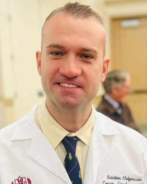
Kristian Holgersson, BS
Loma Linda University School of Medicine
Loma Linda, CA
Presenter(s)
K. Holgersson, Y. Nie, and R. W. Schulte; Loma Linda University, Loma Linda, CA
Purpose/Objective(s): The RTOG0617 trial revealed significantly less survival in patients receiving 74 Gy (experimental arm) compared to those receiving 60 Gy (standard arm) for patients treated with chemoradiation for inoperable stage III non-small cell lung cancer (NSCLC). Post-trial analyses have suggested increased cardiac toxicity as a potential cause of death after dose escalation. We hypothesize that lactic acidosis may contribute to cardiac toxicity due to increased tumor glycolysis in the high-dose arm of 74 Gy. The objective of this study is to evaluate this hypothesis in vitro using murine cardiac and tumor cell lines. Materials/
Methods: Murine cardiac fibroblasts and coronary artery endothelial cells were exposed to x-ray LINAC radiation (6 MV photons), cisplatin, and L-(+)-lactic acid at increasing doses and concentrations to assess if these factors have an additive or synergistic effect of reducing proliferation using MTT assay and Ki67 staining. In addition, post-radiation response (0 Gy, 4 Gy) of Lewis lung carcinoma (LLC1) cells was assessed by measuring L-(+)-lactic acid concentrations in the medium and performing a real-time ATP rate seahorse assay to measure glycolytic vs mitochondrial ATP production rates 48 hours post-irradiation. Cisplatin (0 uM, 5 uM) and glycolysis inhibitor TEPP-46 (0 uM, 400 uM) were added 24 hours before radiation. This was a 2x2x2 factorial design, including negative controls with all combinations tested. Multi-way ANOVA with Bonferroni adjustment for post-hoc analysis was used for statistical analysis (a = 0.05). Additional co-culture experiments with the cardiac cell lines and LLC1 cells are planned to test for interaction between tumor treatment and cardiac cell response.
Results: Results to date revealed a significant additive effect of radiation and L-(+)-lactic acid on proliferation of cardiac cells, with coronary artery endothelial cells being more sensitive than cardiac fibroblasts. The first experiment with the seahorse assay showed a significant reduction of the initially high ATP production rate before treatment, following exposure to radiation and adding TEPP-46 to the medium. TEPP-46, when combined with radiation, normalized the ratio of the high glycolytic relative to the mitochondrial ATP production rate. Additional experiments are needed to confirm these results.
Conclusion: Our in vitro experiments show that radiation and L-(+)-lactic acid have an additive effect on murine cardiac fibroblasts and coronary artery endothelial cells. Radiation combined with TEPP-46 has a strong anti-glycolytic effect on LLC1 cells. This could lead to a reduction of systemic production of L-(+)-lactic acid by lung tumors during treatment and eventually to fewer cardiac side effects in stage III NSCLC patients treated with combined chemoradiation.
